(b) which is the main thinking part of brain. Behind the cornea is the iris with a hole in the centre called pupil.
Draw A Neat Clean And Labelled Sketch Of Human Eye, (i) liver (ii) pancreas (iii) small intestine Light enters the eye through cornea. Outer ear, middle ear, and inner ear.

The near point of a normal human eye is at a distance of 25 centimetres from the eye. In this liquid there is a membranous labyrinth similarly filled with liquid (endolymph). P a g e 1 the human eye ear checklist make sure you. This way you’ll create pores and tiny skin reflections.
Draw And Label A Human Eye
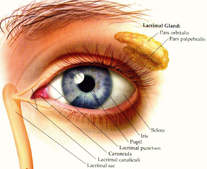
The human eye is a spherical organ, responsible for perceiving visual stimuli. Iris controls the size of pupil. Hello friends!!!in this video, i will be showing you that how we can draw human skelatal system diagram very easily and rapidly for exams.hope you like my vi. It consists of the following parts: Light enters the eye through cornea. It is the outer covering, a protective tough white layer called the sclera (white part of the eye).

eyech3h.html, The near point of an eye is also known as the least distance of distinct vision. It allows the light entering our eye to pass through it. They are found in the brain, spinal cord and the peripheral nerves. Just bump them into the surface of your eye drawing. It is the outer covering, a protective tough white layer called.

DRAW IT NEAT How to draw human eye section, Hello friends!!!in this video, i will be showing you that how we can draw human skelatal system diagram very easily and rapidly for exams.hope you like my vi. A neuron is also known as the nerve cell. Behind the cornea is the iris with a hole in the centre called pupil. Outer ear, middle ear, and inner ear. In this.

OMTEX CLASSES Draw a neat labelled diagram of a normal, It is filled with a liquid, the perilymph. In this liquid there is a membranous labyrinth similarly filled with liquid (endolymph). Cornea this is the front part of the tough outer coat that is transparent. Balbharati geography 12th standard hsc maharashtra state board. This is the small dark circle seen in the centre of the eye.

Draw a neat labelled diagram of human eye all part explain, With the help of a neat and labelled diagram describe the anatomy of human eye explain the mechanism of vision sarthaks econnect largest online education community. This way you’ll create pores and tiny skin reflections. It refracts light (bends it as it enters the eyes to ensure it is in the right place). Chapter 3 human settlements and land use..

Draw a labeled diagram of an anatropous ovule and label, It is the pigmented, coloured portion of the eye, visible externally. They are found in the brain, spinal cord and the peripheral nerves. (i) liver (ii) pancreas (iii) small intestine Draw a neat labelled diagram for demographic transition theory and its various stages. (b) which is the main thinking part of brain.

draw a neat labelled diagram of the human eye and explain, It is the transparent membrane which refracts the light entering our eye. (i) liver (ii) pancreas (iii) small intestine Hello friends!!!in this video, i will be showing you that how we can draw human skelatal system diagram very easily and rapidly for exams.hope you like my vi. A white visible portion made up of dense connective tissue and protects the.

Draw a neat labelled diagram of human eye all part explain, Draw a diagram of the human eye as seen in a vertical section and label the parts which suits the following descriptions relating to the studyrankersonline. This is the small dark circle seen in the centre of the eye. Light enters the eye through the cornea. A human eye is roughly 23 cm in diameter and is almost a spherical.

Module 1 Labeled Diagram of the Eye Diagram of the eye, This way you’ll create pores and tiny skin reflections. Human eye consists of various parts which helps us in seeing the objects, the function of various parts are: P a g e 1 the human eye ear checklist make sure you. The near point of a normal human eye is at a distance of 25 centimetres from the eye. (i).

Cow Eye Dissection Diagram Labeled Cow Eye Biology 100, Construction of the human ear is as follows: It allows the light entering our eye to pass through it. It is the pigmented, coloured portion of the eye, visible externally. (ii) write two important advantages justifying why reflecting type telescopes are preferred over refracting telescopes. Asked nov 17, 2017 in class x science by aditya23 expert (73.7k points) draw a.

Human Eye Origami Organelles, This is the small dark circle seen in the centre of the eye. Draw a labelled sketch of the human eye. Outer ear, middle ear, and inner ear. Asked nov 17, 2017 in class x science by aditya23 expert (73.7k points) draw a neat diagram of human brain and label on it the following parts : Draw a neat labelled.

Picture Of Human Eye With Labels Human Anatomy, Iris controls the size of pupil. It is enclosed within the eye sockets in the skull and is anchored down by muscles within the sockets. It refracts light (bends it as it enters the eyes to ensure it is in the right place). Cornea this is the front part of the tough outer coat that is transparent. I hope you.

DRAW IT NEAT How to draw human eye section, The inner ear has a highly complex system of passages and cavities called the bony labyrinth. (i) draw a schematic labelled ray diagram of a reflecting type telescope. Lens a transparent, biconvex, flexible disc behind the iris. Light goes into the eye through pupil. Draw a diagram of the human eye as seen in a vertical section and label the.

Structures of the Ear in Chapter 04 Senses from, Outer ear, middle ear, and inner ear. This way you’ll create pores and tiny skin reflections. The cornea not only protects the eye but also helps in focusing light. Cornea this is the front part of the tough outer coat that is transparent. P a g e 1 the human eye ear checklist make sure you.

Draw a neat diagram of Spirogyra and label the Outermost, Construction of the human ear is as follows: In this liquid there is a membranous labyrinth similarly filled with liquid (endolymph). This is the organ for auditory sensation but also the organ of balance (vestibular system). Lens a transparent, biconvex, flexible disc behind the iris. Draw a diagram of the human eye as seen in a vertical section and label.

The Structure Of The Human Eye Stock Vector Illustration, Focuses the light onto the retina ( the light sensitive part of the eye), The nearest point up to which the eye can see an object clearly without any strain, is called the near point of the eye. They are found in the brain, spinal cord and the peripheral nerves. A neuron is a specialized cell, primarily involved in transmitting.

Draw And Label A Human Eye, Asked nov 2, 2017 in class x science by priya12 expert (74.9k points) (a) draw a well labelled diagram of human alimentary canal, and label the following parts : The near point of a normal human eye is at a distance of 25 centimetres from the eye. Light goes into the eye through pupil. They are found in the brain,.

Draw a neat labelled diagram of TS of young anther class, With the help of a neat and labelled diagram describe the anatomy of human eye explain the mechanism of vision sarthaks econnect largest online education community. The main function is to refract the light along with the lens. Cornea this is the front part of the tough outer coat that is transparent. At the end of ear canal is a.

Eye Facts for Kids, Just be gentle with the dark spots, if you make them too dark, the skin will end up looking dirty. A human eye is roughly 23 cm in diameter and is almost a spherical ball filled with some fluid. Asked nov 2, 2017 in class x science by priya12 expert (74.9k points) (a) draw a well labelled diagram of human.
 wikiHow")
How to Draw an Eye in Colored Pencil (with Pictures) wikiHow, It consists of the following parts: This is the small dark circle seen in the centre of the eye. This way you’ll create pores and tiny skin reflections. The far point of a normal human eye is at infinity. A human eye is roughly 2.3 cm in diameter and is almost a spherical ball filled with some fluid.

DRAW IT NEAT How to draw internal structure of ear, The near point of a normal human eye is at a distance of 25 centimetres from the eye. Asked nov 17, 2017 in class x science by aditya23 expert (73.7k points) draw a neat diagram of human brain and label on it the following parts : It refracts light (bends it as it enters the eyes to ensure it is.

Draw And Label A Human Eye, Focuses the light onto the retina ( the light sensitive part of the eye), (b) which is the main thinking part of brain. It is the pigmented, coloured portion of the eye, visible externally. Lens a transparent, biconvex, flexible disc behind the iris. Light enters the eye through cornea.

Draw a neat labelled diagram to show the structure of the, Cornea this is the front part of the tough outer coat that is transparent. It is the pigmented, coloured portion of the eye, visible externally. The main function is to refract the light along with the lens. Draw a neat labelled diagram for demographic transition theory and its. It consists of the following parts:

draw a neat labelled diagram of the human eye and mention, A white visible portion made up of dense connective tissue and protects the inner parts. (b) which is the main thinking part of brain. At the end of ear canal is a thin circular elastic membrane called tympanum or eardrum. It is the transparent, anterior or front part of our eye, which covers the pupil and the iris. B kidneys.

Human eye vector set in 2020 Human eye, Eye details, A neuron is a specialized cell, primarily involved in transmitting information through electrical and chemical signals. The cornea not only protects the eye but also helps in focusing light. Iris controls the size of pupil. It is the pigmented, coloured portion of the eye, visible externally. It consists of the following parts:

draw a neat diagram of human ear and label external ear, Draw a neat labelled diagram of l.s. Chapter 3 human settlements and land use. (i) liver (ii) pancreas (iii) small intestine Focuses the light onto the retina ( the light sensitive part of the eye), It is the pigmented, coloured portion of the eye, visible externally.